Издательство: ИПК СВФУ
Год выпуска: 2011
Количество страниц: 142 с.
Издательство: ЯГУ
Год выпуска: 2001
Количество страниц: 68 с.
Ответственность: Чугунова Саргылана Афанасьевна (Составитель), Николаева Татьяна Яковлевна (Составитель), Тырышкина Эржена Васильевна (Составитель), Горохова Олесия Алексеевна (Составитель), Конникова Эдилия Эдуардовна (Прочие), Кудрина Полина Ивановна (Прочие)
Год выпуска: 2018
Количество страниц: 52 с.
Methodical recommendations are intended for neurologists, neurosurgeons, endovascular surgeons, clinical residents, graduate students and students of medical universities
Алгоритмы диагностики острых нарушений мозгового кровообращения : методические рекомендации / [составители: кандидат медицинских наук С. А. Чугунова, доктор медицинских наук Т. Я. Николаева, Э. В. Тырышкина, О. А. Горохова]. – Якутск : [б. и.], 2018. – 47 с.
Количество страниц: 6 с.
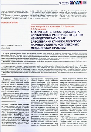
Анализ деятельности кабинета когнитивных расстройств центра нейродегеративных заболеваний клиники Якутского научного центра комплексных медицинских проблем / Ю. И. Хабарова, З. Н. Алексеева, Т. К. Давыдова, О. В. Татаринова // Якутский медицинский журнал. — 2020. — N 3 (71). — С. 119-124. – DOI: 10.25789/YMJ.2020.71.30.
DOI: 10.25789/YMJ.2020.71.30
Количество страниц: 4 с.
100 villous chorions at 9-12 weeks of pregnancy were studied in order to identify the relationship between the indicators of progesterone synthetic activity of the fetoplacental complex being formed and the nature of clinical and echographic manifestations of the threatened miscarriage in groups of women with different course of cytomegalovirus infection (CMV) infection. It was identified a significant dependence of the frequency of the threatened miscarriage and its clinical and echographic manifestations on the exacerbation of CMV infection in the first trimester of pregnancy, which was combined with low values of progesterone and 3β-hydroxy-5-pregnen-20-one-dehydrogenase in the villous chorion compared to the group with a latent course of infection
Никитина, М. А. Апатия как немоторный симптом болезни Паркинсона и болезни Гентингтона / М. А. Никитина, Н. Г. Бразовская, Н. Г. Жукова [и другие] // Якутский медицинский журнал. — 2020. — N 2 (70). — С. 24-27. – DOI: 10.25789/YMJ.2020.70.07.
DOI: 10.25789/YMJ.2020.70.07
Количество страниц: 8 с.
The article presents the results of a clinical and genetic study of a Yakut family with hereditary spastic paraplegia (HSP). Patients with clinically diagnosed HSP and healthy family members were studied. The disease is clinically characterized as a progressive spastic paraplegia of the lower extremities concomitant peripheral neuropathy in advanced case. The methods of exome sequencing of the entire genome, molecular modeling of dynamin-2 and experimental reproduction of key elements of the HSP pathogenesis have been applied. Genetic analysis revealed a novel missense c.2155C> T, p.R719W mutation in the highly conserved GTP-effector domain of the dynamin-2 gene (DNM2). In experiments on HeLa cells, it was shown that mutant dynamin-2 affected endocytosis process. In-silico modeling determined that the identified mutation is located in the DNM2 bundle-signaling element and potentially disrupts the assembly and functional properties of the protein. Testing of this mutation in other Yakut families with HSP showed a negative result, which once again confirms the genetic heterogeneity of this pathology
Аутосомно-доминантная спастическая параплегия в четырех поколениях якутской семьи, вызываемая мутацией в динамине-2 / Т. М. Сивцева, Л. Г. Гольдфарб, Т. К. Давыдова [и другие] // Якутский медицинский журнал. — 2020. — N 1 (69). — С. 6-12.
DOI: 10.25789/YMJ.2020.69.01
Количество страниц: 4 с.
To date, there are no methods of treatment and prevention of the development of epilepsy in people at risk. All this indicates the need for a search for biomarkers of epileptogenesis, diagnosis, disease progression, drug response and treatment safety. As biomarkers of epilepsy, the following are considered: electrophysiological changes, the presence of a clinical attack, genetic changes, micro ribonucleic acid (microRNA) of plasma / serum / cerebrospinal fluid; protein biomarkers, plasma exosome biomarkers, cerebral cortex microRNAs; biomarkers, strain gauge images / diffusion-weighted images of magnetic resonance imaging (MRI). The authors review the literature on modern studies of various biomarkers of epilepsy, which allow a personalized approach to assessing the diagnosis, treatment and response to epilepsy therapy.
Биомаркеры эпилепсии / К. Д. Яковлева, М. Р. Сапронова, А. А. Усольцева, Ю. С. Панина, С. Н. Зобова, Д. В. Дмитренко // Якутский медицинский журнал. — 2019. — N 4 (68). — С. 99-102.
DOI: 10.25789/YMJ.2019.68.28
Количество страниц: 6 с.
- Математика. Естественные науки > Общая биология. Антропология. Вирусология. Микробиология,
- Прикладные науки. Медицина. Ветеринария. Техника. Сельское хозяйство > Медицина > Патология. Клиническая медицина > Неврология,
- НАУКА ЯКУТИИ > ПРИКЛАДНЫЕ НАУКИ. МЕДИЦИНА. ТЕХНИКА. СЕЛЬСКОЕ ХОЗЯЙСТВО > Медицина > Патология. Клиническая медицина > Неврология,
- НАУКА ЯКУТИИ > МАТЕМАТИКА. ЕСТЕСТВЕННЫЕ НАУКИ > Общая биология. Антропология. Вирусология. Микробиология.
This article considers the use of microRNAs as a possible biomarker of epilepsy. The presented studies have shown that microRNAs can be involved in the process of epileptogenesis by regulating the inflammatory response, apoptosis of neurons, and transcription factors involved in cell differentiation. Biological fluids (blood and CSF) of patients with epilepsy showed differences in the number of circulating microRNAs, which may allow further use of microRNAs as a diagnostic biomarker. Recent discoveries providethe sufficient source of new microRNA targets, but there are still significant problems of studying their role in pathogenesis and the possibility of their application in clinical practice
Биомаркеры эпилепсии: микроРНК / М. Р. Сапронова, К. Д. Яковлева, А. А. Усольцева [и другие] // Якутский медицинский журнал. — 2020. — N 4 (72). — С. 106-111
DOI: 10.25789/YMJ.2020.72.26
Количество страниц: 4 с.
Тихонов Д. Г., Вероятные причины вилюйского энцефаломиелита. Факты истории изучения и размышления // Якутский медицинский журнал. 2019. – N 1 (65). – С. 115-118.
DOI: 10.25789/YMJ.2019.65.35
Количество страниц: 6 с.
Parkinson’s disease and essential tremor are the most common neurodegenerative diseases, which are accompanied by the development of tremor - involuntary rhythmic hyperkinesis. In the essential tremor,tremor has a kinetically postural character with a frequency of 6 to 10 Hz, affectingarms, head, face, vocal cords, tongue, legs and trunk. Parkinson tremor has a frequency of 4-6 Hz, occurring at rest in the form of “counting coins” or “rolling pills”, decreasing with active movements until complete disappearance, and is characterized by resumption after 2 or more seconds after giving the pose (“renewed” tremor). The generation of tremors is associated with dysfunction of two main ways: basal nuclei - cerebellum - thalamus and cogwheel - olive complex. Consequently, damage to any of these levels leads to the development of tremors. Recently, the concept of a single continuum of essential tremor - Parkinson’s disease - is widely discussed.It has been established that these neurological disorders can coexist or transit into each other. So, essential tremor increases the risk of developing Parkinson’s disease by 3.5 times, and in patients with Parkinson’s disease the likelihood of attaching an essential tremor is as high. This is due to the cross pathophysiological mechanism, which is probably due to genetic mutations. This article discusses the pathophysiological mechanisms of the development of tremor in patients with Parkinson’s disease and essential tremor. We describe a patient with the essential tremor whose disease has been transformed into tremor predominant Parkinson’s disease
Таппахов, А. А. Взаимосвязь болезни Паркинсона и эссенциального тремора: обзор литературы и клинический случай /А. А. Таппахов, Т. Г. Говорова, Т. Е. Попова // Вестник Северо-Восточного федерального университета им. М. К. Аммосова. Серия: Медицинские науки.— 2018. — N 2 (11). — С. 44-49.